More Information on Suture Closure

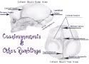
The Skull of an Infant is made up of bones that are separated by fibrous joints (sutures). In a normal skull not affected by craniosynostosis these fibrous joints remain open and flexible during the child's first years of life to expand and grow with the rapid growth of the infant's brain.
If one or more of these fibrous sutures closes to early the skull cannot expand normally with growth of the brain, and so assumes an abnormal shape: Normocephaly(Skull with no Craniosynostosis)

Metopic Craniosynostosis:

The metopic suture begins at the nose and continues to meet the sagittal suture. Metopic craniosynostosis results in a narrow, triangular forehead with pinching of the temples laterally(trigonocephaly).
The Metopic Suture is the first and only suture which normally fuses early in childhood(normally around 6 to 8 mths but in some cases as early as 3 months.) Metopic Craniosynostosis is one of the least common forms of Craniosynostosis. However, sometimes the early fusion of the Metopic suture will cause a Metopic ridge with no trigonocephaly. In cases of a metopic ridge surgical intervention may not be needed.
In true Metopic Craniosynostosis with the evidence of trigonocephaly (a triangular forehead and narrowing to the skull), whether this narrowing is mild or severe, many experienced surgeons will recommend that the infant's skull should be surgically repaired.
If your child has a Metopic ridging with no trigonocephaly be sure to consult a craniofacial specialist. Even if you are advised to be wait and see always research and ask questions.
Saggital Craniosynostosis:

The sagittal suture runs from the front of the head to the back of the skull. Fusion of the suture results in a long, narrow skull which may or may not include bulging of both the front and back of the head. The shape of a skull affected with Saggital Craniosynostosis is also known as scaphocephaly. Saggital Craniosynostosis is the most Common form of and estimated to happen one in 1000 births.
It is thought that disproportion in the size of the pelvis of the mother and the size of the fetal head can cause Saggital Craniosynostosis. The limited room for growth is thought to bring the the two parietal bones together too early creating the synostosis in the pelvis before birth. This is why Saggital Craniosynostosis is often seen in first pregnancies. Also, boys have larger heads and that is why it is thought more boys than girls are affected with Saggital Synostosis.Even with these thoughts on Saggital Craniosynostosis there is nothing that an expectant mother can do to prevent craniosynostosis. There is still not enough known about Saggital and any other form of Craniosynostosis. Research is still being discovered for the causes of premature suture closure.
Coronal Synostosis:
The coronal sutures begin at the ear and continue superiorly to the top of the skull to meet the sagittal suture. One or both sutures may be involved.
When one suture is fused that is known as
Unilateral (left or right) Coronal synostosis

The shape of the head is sometimes referred to as plagiocephaly (which should not be confused with positional plagiocephaly.) The forehead on the effected side is flattened and swept back with the eye in its socket. Unilateral Cranio can give the appearance that one eye is wider open then the other which is caused by the lack of orbit around the affected eye.
When both sutures are fused that is known as bicoronal synostosis.

The head is short and wide with the head shape being referred to as brachycephalic. There is a lack of growth with both eye orbits which can cause bulging of the eyes. The back of the head might appear flat with a bulging of the skull around the ears. The skull tends to tower upwards as it continues to grow.
Coronal Craniosynostosis may or may not be caused by a syndrome. One syndrome babies with Coronal Synostosis can commonly have is Muenke's Syndrome. Along with the coronal suture fusion a few more symptoms of Muenke's may be, flattened cheek bones/shallow mid-face, wide-set eyes, hand or feet abnormalities(my daughter has Muenke's syndrome and the middle bones of her fingers are shorter than normal), possible hearing loss, and possible learning delays. In the cases of multisuture craniosynostosis your Craniofacial Specialist might recommend meeting with genetics for follow-up and testing.
Lambdoid Craniosynostosis:

The Lambdoid suture is located on the back of the skull. It has a right and left side and is shaped like an upside down "V.
One key marker for fusion of the lambdoid suture is a low bump behind the ear on the same side as the fused suture. Another good way to determine if the lambdoid suture is closed is to look and see if the ear on the same side as the posterior skull flatness is pulled backwards and sticks out more. If the ear is forward on the flat side, with respect to the opposite ear, then a skull deformation should be suspected instead of a fused suture. When viewed from above, the affected side of the back of the head is flatter than the opposite side.
Lambdoid Craniosynostosis is the rarest form of Craniosynostosis and fusion of both Lambdoid sutures is more rare. The diagnosis of Lambdoid synostosis is the most difficult to make because on plain skull x-rays the lambdoid suture is frequently misdiagnosed as being fused shut. Ideally, the diagnosis of lambdoid synostosis is made by CT scans, read at an experienced center. Children who have lambdoid synostosis and significant flattening.Cloverleaf Craniosynostosis:

Also known as kleeblattschadel. Premature fusion of the coronal, lambdoid and posterior sagittal sutures results in a cloverleaf skull. This is the rarest form of Craniosynostosis and evidence of closure will be evident at birth. Due to the severity of multiple sutures being fused children with Cloverleaf should be closely followed by pediatric neurosurgeons, pediatric craniofacial surgeons, and team of doctors to follow up through childhood and possibly adulthood.
How is Craniosynostosis Fixed?
There is no cure for Craniosynostosis.
The only treatment is surgery.
Parents that suspect something wrong with their child's head shape should consult with a pediatric craniofacial surgeon and/or pediatric neurosurgeon.
The main purpose for surgery is to open up the skull to allow proper skull growth as well as to correct the shape of the skull. As more of our children are affected with Craniosynostosis research is being done on the impact fused sutures might have on brain function. Some of our children might have cognitive delays while others will grow up with no affects from the synostosis and surgery. This is a scary time for parents of children with craniosynostosis. It opens up a whole roller coaster of emotion. There are groups that will help you face prediagnosis concerns, surgery, and postop care CAPPSKids.org and CranioKids.org as well as the JorgePosada Foundation strive to help families affected with Craniosynostosis. Their links and info are provided on our support page.
Click here for another great site for Craniosynostosis and Positional Plagiocephaly Information is
Go HERE for more information on Types of Surgery for Craniosynostosis
If not Craniosynostosis? What's wrong with my baby's head? Info on Positional Plagiocephaly here